Specialities
Micro-endoscopic Disc Surgery (Minimally Invasive Disc Surgery/Key hole disc surgery)
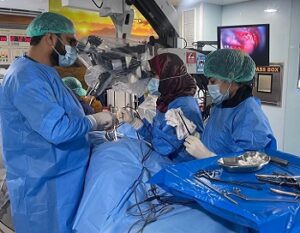
Micro-Endoscopic Disc surgery also known as Key Hole disc surgery, is a recently introduced surgical technique in Pakistan for treatment of prolapsed intervertebral disc. It is famous procedure all over the world, for excision of disc fragments causing stenosis or nerve root compression (sciatica) using minimally invasive surgical techniques. This procedure is highly effective in relieving symptoms & results in early recovery from surgery, thus allowing discharge from hospital & mobilisation of patient only few hours after operation. Key Hole disc surgery is also much safer than open surgery & leaves less than 2cm scar, thus having much better cosmetic results.
Endoscopic Brain Tumor Surgery
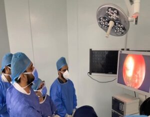
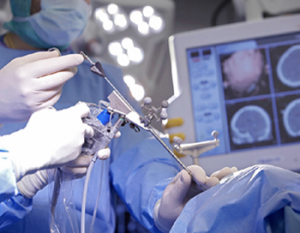
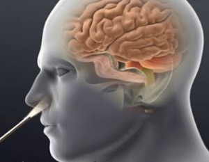
Endoscopic brain surgery is minimally invasive brain surgery used traditionally for Endoscopic third ventriculostomy but it’s use has expanded in last few decades. There are several brain tumors that can excised endoscopically such as Pituitary Tumors, Planum Meningiomas and clival tumors. Similary some tumors of third ventricle can also be excised using minimally invasive endoscopic techniques. The major advantages of brain endoscopy include minimum damage to normal structures, no brain retraction, better panoramic vision & less blood loss. Therefore, endoscopic surgery (where possible) is preferred over open surgery in modern Neurosurgical era.
Deep brain stimulation for Parkinsons, Dystonia & Esential tremors
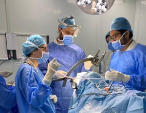
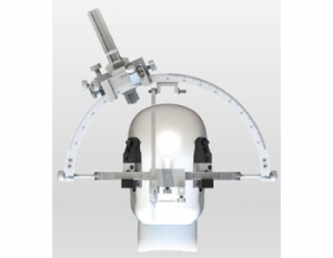
Deep Brain Stimulation is highly advanced, sophisticated & safe, minimally invasive brain surgery. The fine electrodes ( DBS leads) are implanted in specific areas of brain to get desired effects & control of symptoms. These DBS leads are then connected to battery placed in upper part of front chest. The delivery of electric current from battery to brain leads is controlled with programmer & can be adjusted to individual patient needs. Most of this surgery is performed while patient is awake & noticing the effects of stimulation & giving feedback how he is feeling with therapy. DBS therapy is approved by FDA for treatment of Parkinsons Disease, Dystonia & Essential tremors. Although the DBS treatment is expensive but it brings marked improvement in patient’s quality of life.
Minimally Invasive Spinal Fixation for Spinal Injuries
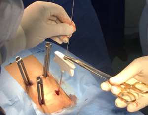
Minimally invasive spinal fixation or Percutaneous spinal fixation is latest technique used to treat patients with spinal fractures of thoracic & lumbar spine. It is latest technique that circumvented the need of large skin incisions & extensive tissue dissection for stabilising the spine. The sophisticated technique needs very small skin incisions , retraction of muscles with minimal disruption & placement of screws in spine (vertebral body) under image guidance.In contrast to traditional spinal fixation, minimal invasive spinal fixation has much less tissue damage & better safety profile, therefore is much preferred over traditional fixation techniques.
Endoscopic Third Ventriculostomy
Hydrocephalus is a Neurosurgical condition that results from impaired absorption or circulation of Cerebrospinal fluid (CSF) in brain. Congenital abnormalities, infections & trauma are among commons causes of Hydrocephalus. The increased volume of CSF in brain leads raised Intrcranial pressure, thus can be life threatening condition. Traditionally, the treatment of Hydrocephalus was done with diversion of CSF from brain to abdomen using Shunt devices, however it’s associated with several risks, the most feared are infection and malfunction. In modern day Neurosurgery shunts are replaced with Endoscopic Third ventriculostomy, in which minimal invasive brain surgery techniques are used to make small opening in floor of Ventricle, thus allowing CSF absorption in natural way, by bypassing the obstruction in CSF pathway. The procedure obviates the need of Abdominal incision needed for VP shunt. Therefore, Endoscopic Third Ventriculostomy is safe & effective procedure, as no hardware is implanted in brain & absorption of CSF takes place by natural means.
Cervical Spine Fixation
Injuries of cervical spine are quite common & leading cause of disability in our country. The timely surgery of spinal injuries can help in recovery from disability & many patients can regain their movements. Fixation of cervical spine is delicate surgery that requires knowledge & skill. It can be done front or back of neck, depending on the type disease/injury.
Anterior cervical fixation is done with small incision in-front of neck & most of the procedure is done under microscope. The disc or vertebral body is removed & replaced with special implants, followed by screws & plates to secure implant at it’s place. The recovery from surgery is quick & patients can be discharged from hospital on next day of surgery.
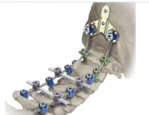
Posterior cervical fixation is done through incision at back of neck. The muscles are dissected from vertebrae, screws are placed in vertebra & connected with rods. This provides rigid support of cervical spine (neck). Like Anterior fixation, patients of posterior spinal fixation can also be discharged from hospital one day after surgery.
Craniocervical Junction Fixation
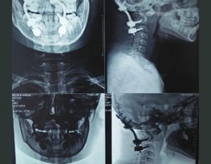
The junction of Skull ( head )with Spine is complex, as large number of ligaments & special vertebrae are involved in providing wide range of mobility & maintaining spinal stability. Wide range of diseases like infection, trauma, tumours & arthritis, can lead to bony destruction or ligamentous disruption at craniocervical junction, rendering it unstable & resulting in pain & weakness of limbs. In most cases surgical stability is required which is achieved by fixation through screws and rods. The complex anatomy at this craniocervical junction, makes surgery challenging & high precision is required to achieve desired results without devastating complications. Dr. Zubair Mustafa Khan has successfully performed large number of such complex surgeries with excellent outcome.
Endoscopic CSF Rhinorrhea Repair
The human brain is surrounded by a clear fluid called Cerebrospinal Fluid (CSF), which protects brain from injury, provides nutrients to brain & helps in removal of unwanted materials from brain. In some pathologic conditions like brain tumours, hydrocephalus or after head injury, there is leakage of CSF from nose. This can lead to serious life threatening conditions like meningitis, tension pneumocephalus or intracranial haematoma. Fortunately most of CSF leaks stop spontaneously but in cases where it persists, it should be treated appropriately to prevent serious complications. Traditionally CSF leakage has been treated through open brain surgery but now most of the cases of CSF Rhinorrhea can be treated endoscopically with more than 90% success rate. The procedure is done through nose thus no incision is required & complications of open brain surgery are also avoided.
